Positive Urine Cytology Reveals Melanoma
Rare case links prior melanoma in situ to ureter and porta hepatis metastases
A 77-year-old woman developed metastatic melanoma four years after excision of a lesion previously diagnosed as melanoma in situ. Presenting with symptoms like hematuria and abdominal pain, imaging revealed extensive metastases primarily affecting the right ureter and porta hepatis. Histopathological evaluations confirmed metastatic disease, distinguishing it from primary urothelial carcinoma. The case highlights the rarity of such occurrences and emphasizes the critical need for thorough assessment and immunohistochemical analysis in atypical genitourinary tumors, ensuring accurate diagnosis and prompt treatment with systemic immunotherapy.
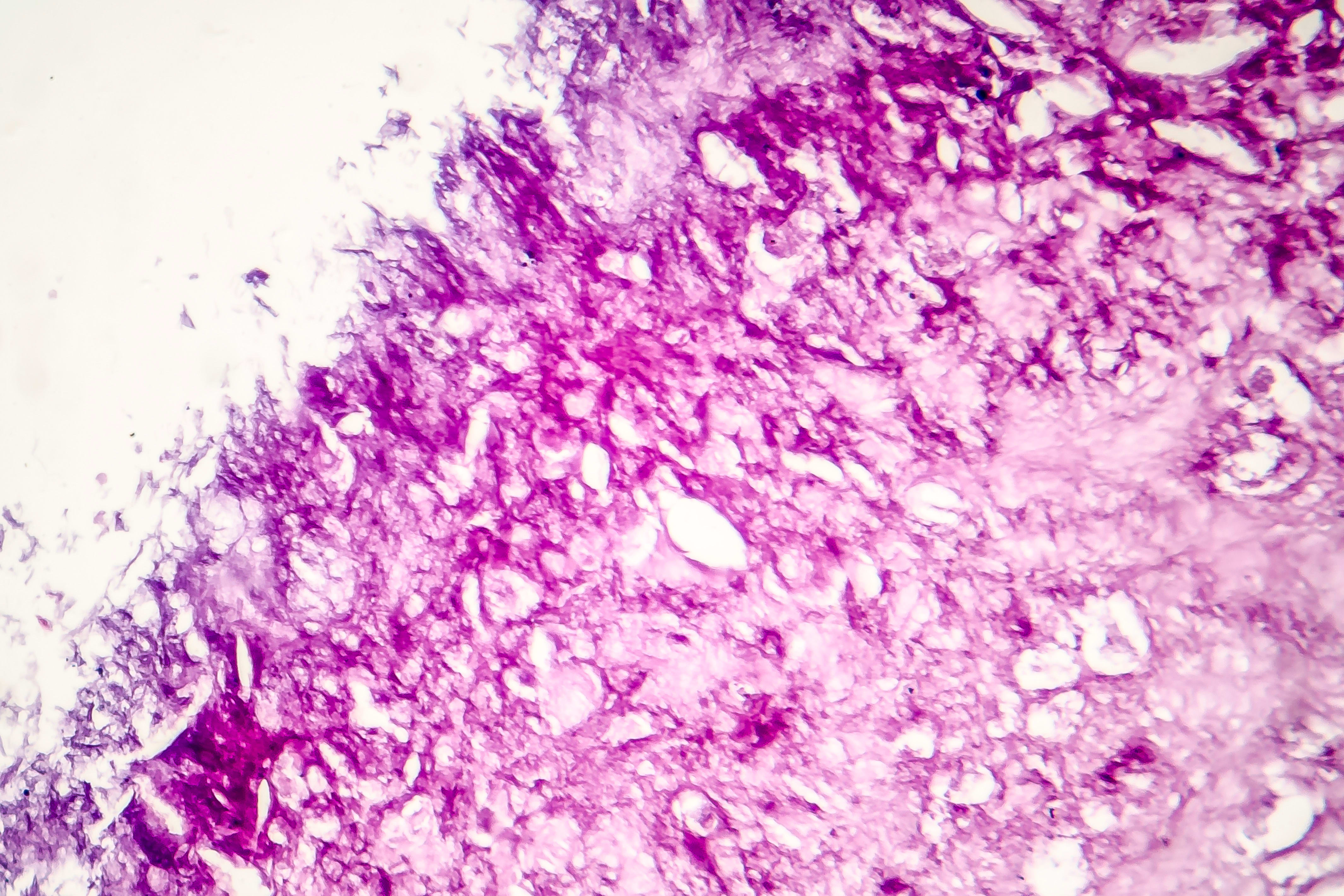
1. Metastatic melanoma can occur years after melanoma in situ excision.2. Atypical genitourinary lesions should include metastatic melanoma in differential diagnosis.3. Immunohistochemical evaluation is critical for accurate diagnosis.4. Ureteral involvement in melanoma is rare (0.2% to 1%).5. Patient was treated with pembrolizumab after diagnosis of metastatic melanoma.