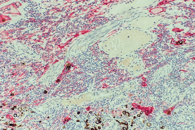
A keratin/D2-40 dual stain was used on a colon resection specimen to visualize lymphovascular invasion by tumor cells. The staining technique highlights lymphovascular spaces in red, while the keratin-positive carcinoma cells appear brown, facilitating the identification of cancer spread within the vascular system. This methodology is crucial in assessing the aggressiveness of colorectal tumors and their potential to metastasize, impacting treatment decisions.
1. Keratin/D2-40 dual stain aids in detecting lymphovascular invasion. 2. Red highlights lymphovascular spaces; brown indicates carcinoma cells. 3. Essential for assessing tumor aggressiveness. 4. Helps determine potential for cancer spread (metastasis). 5. Important in colorectal cancer treatment planning.
Listen Tab content